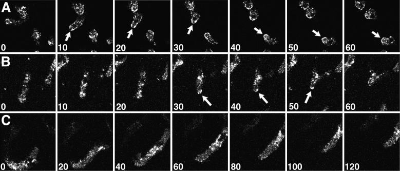
Figure 6.
Clathrin localizes to the tail of moving cells. Confocal images were collected from fields of Dictyostelium cells expressing GFP-clathrin during cell locomotion; the numbers indicate the time interval (seconds) between frames. (A and B) During tail retraction, translocating cells displayed enriched GFP-clathrin at their posterior edges (arrows). (C) A cell translocating without tail retraction showed no enrichment of GFP-clathrin in its tail. See accompanying video. Video is 80 times real time.

