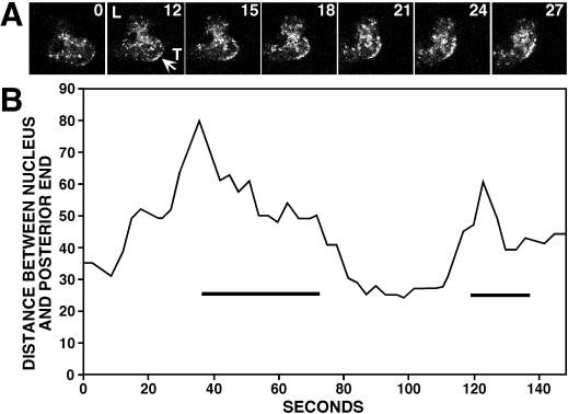
Figure 7.
Clathrin localization to the posterior plasma membrane during tail retraction. (A) Time sequence taken of a translocating cell. The numbers indicate the time interval (seconds) between frames. GFP-clathrin concentrated at the posterior plasma membrane coincident with tail retraction (arrow). (B) The distance between the nucleus and the posterior end of a typical cell undergoing two cycles of lamellipodia extension and tail retraction was measured from confocal images taken every 3 s. GFP-clathrin localization to the posterior plasma membrane (bars) coincided with tail retraction.

