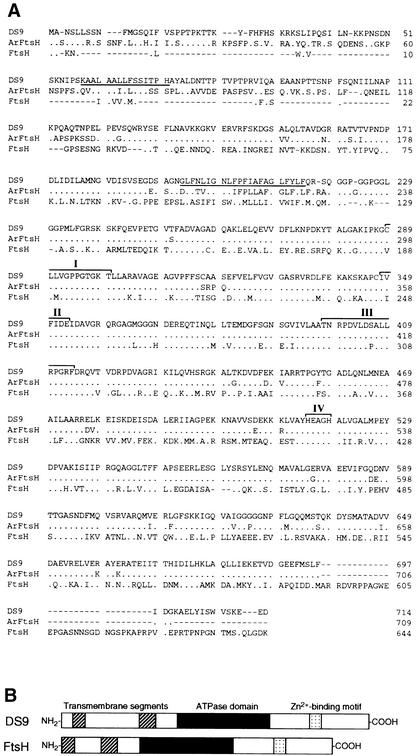
Figure 2.
Comparison of the Deduced Amino Acid Sequences of DS9, Arabidopsis FtsH Homolog, and E. coli FtsH.
(A) Lines at top, center, and below represent the amino acid sequence of DS9 (GenBank accession number AB017480), an Arabidopsis FtsH homolog (Lindahl et al., 1996), and E. coli FtsH (Tomoyasu et al., 1993a), respectively. The underlined amino acids represent putative membrane-spanning regions. Dots represent identical amino acid residues, and dashes indicate gaps introduced to maximize alignment. The roman numerals indicate two regions of ATP binding motif (I and II), a second region of homology(III), and a Zn2+ binding motif (IV).
(B) Diagrammatic alignment of the primary structures of DS9 and E. coli FtsH. Hatched, black, and stippled boxes represent two putative transmembrane segments, the ATPase domain, and the putative Zn2+ binding motif, respectively. The ATPase domain contains an ATP binding motif and a second region of homology (Confalonieri and Duguet, 1995). The putative Zn2+ binding motif is HEXXH, where X indicates nonconserved amino acid residues.

