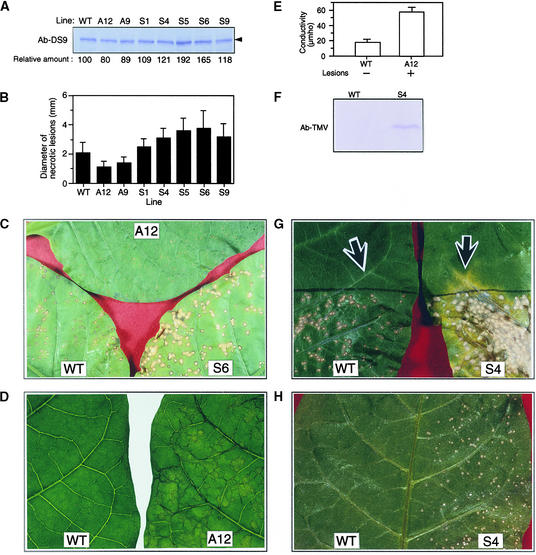
Figure 8.
Analysis of Transgenic Tobacco Plants Containing Various Amounts of the DS9 Protein.
The upper, fully expanded healthy leaves of 3-month-old wild-type Samsun NN (WT) and transgenic tobacco plants (S4, S6, and A12) were used for each experiment.
(A) Total protein was extracted from the wild-type and transgenic tobacco plants carrying antisense (lines A12 and A9) and sense (lines S1, S4, S5, S6, and S9) constructs. Twenty-five micrograms of total protein from each line was subjected to protein gel blot analysis with antibodies raised against DS9ΔN (Ab-DS9). The numbers below the gel indicate the amount of DS9 protein expressed as a ratio of that present in the wild type, which was set equal to 100%. The arrowhead indicates the relative molecular masses of ∼78 kD.
(B) Leaves were inoculated with TMV (2 μg/mL) and incubated at 20°C under 120 μmol of photons m−2 sec−1 fluorescence illumination. In each line, the diameter of 60 local lesions 5 days after inoculation was measured with a stereoscopic microscope. Each bar represents the mean ±sd.
(C) Necrotic lesions that had formed on the leaves of a wild-type plant and the S6 and A12 lines, as described in (B).
(D) Leaves of line A12 that had been inoculated with TMV (10 μg/mL) and incubated for 40 hr at 30°C were shifted to 20°C, where they were incubated for 3.5 hr and returned to 30°C. As a control, leaves of wild-type plants were used. A photograph was taken 24 hr after returning the leaves to 30°C. The experiment was repeated twice with similar results.
(E) After photographing the leaves as shown in (D), five leaf discs were punched from each leaf and measured for electrolyte leakage. Values are the mean ±sd from three independent experiments. +, lesions appeared; −, no lesions appeared.
(F) Protein gel blot analysis of TMV coat protein. The experiment was repeated twice with similar results. Ab-TMV, anti-TMV antibody.
(G) The bottom half of leaves of wild-type and S4 plants were inoculated with TMV (2 μg/mL) and incubated at 20°C. Seven days after inoculation, the leaves were photographed. Two discs (0.7 cm in diameter each) from each leaf were punched out from the sites indicated by the arrows,
which were 1.5 cm away from the black borderline that indicates the edge of the inoculated region. One of the two discs from each leaf was used for protein gel blot analysis with the anti-TMV antibody (F), and the other was used for the TMV bioassay (H).
(H) TMV bioassay. For the TMV bioassay, crude leaf extracts from wild-type plants and the S4 line were inoculated onto the left and right half, respectively, of Samsun NN tobacco leaves. Five days after inoculation, the leaf was photographed. The experiment was repeated twice with similar results.

