Figure 7.
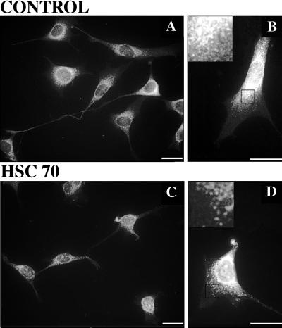
Exogenous hsc70 releases kinesin from gently permeabilized cells. Kinesin immunofluorescence in untreated (A and B) and hsc70-treated (C and D) cells is shown. Images A and C were acquired with a 40×, objective and B and D were acquired with a 63× objective; calibration bars are 20 μm in each case. Insets in B and D were digitally enlarged (4×) from the areas demarcated by black boxes. Kinesin inmunoreactivity declined after hsc70 treatment. Comparing A and C, the amount of kinesin in processes and in the perinuclear halo is reduced. Comparing B and D, this erosion of kinesin immunoreactivity in the perinuclear halo is more readily visulalized. The effect is most obvious near the cell center (B and D, insets), where the density of punctuate structures is reduced, resulting in a thinner perinuclear stain. Longer treatments will remove more kinesin immunoreactivity.

