
Figure 5.
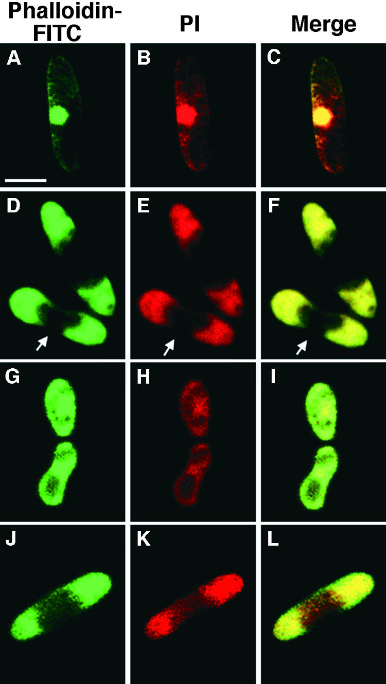
Confocal Microscopic Analysis of Germinating CgMEK1 Mutant Conidia and MEK Inhibitor–Treated Wild-Type Conidia.
(A) to (I) CgMEK1 mutant conidia were incubated on glass slides for 0 hr ([A] to [C]), 12 hr ([D] to [F]), or 24 hr ([G] to [I]) at a low population density (10 conidia per μL) in the absence of host chemical signals.
(J) to (L) Wild-type conidia, also at 10 conidia per μL, were incubated on glass slides for 12 hr in the presence of 10 μM PD98059.
Images were visualized by phalloidin-FITC signals (left), PI signals (middle), and merged images (right). Arrows indicate the constriction of the conidium.  .
.