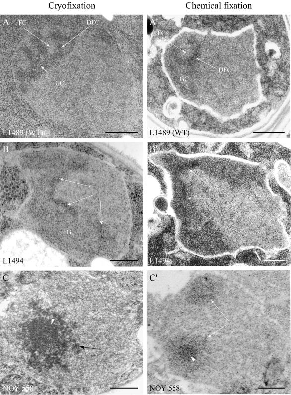
Figure 1.
Nuclear ultrastructure of S. cerevisiae mutants L1494 and NOY 558. Yeast strains L1489 (wild-type), L1494 (rdnΔ, pRDN-wt), and NOY 558 (rrn7Δ, pGAL7-RDN) were cryofixed (A–C) or chemically fixed (A′–C′) and prepared for electron microscopy. The nucleolus in wild-type strain L1489 (A and A′) presented the three typical components: FCs, a DFC, and a GC. In mutant L1494 (B and B′), the nucleus displayed an electron-dark region made of a fibrillar network (arrows) and a granular region (G). The nucleus of mutant NOY 558 (C and C′) contained one or several electron-dark regions, referred to as mininucleoli (arrows). Two domains were identified in mininucleoli: a central compact region (arrowhead) surrounded by a diffuse region presenting mostly a fibrillar aspect. Bar, 300 nm.

