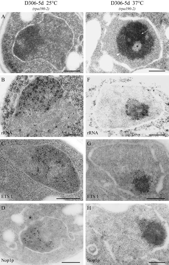
Figure 11.
Electron microscopic analysis of the nucleolar dynamics in strain in D306-5d according to rDNA transcription state. At 25°C, strain D306-5d exhibited a wild-type nucleolus (A). Nuclear rRNA (B), ETS 1 (C), and Nop1p (D) were located in the nucleolus. Upon transfer to 37°C for 4 h, the nucleoli segregated into three major components (E): low-electron-dense domains (asterisk) surrounded by a high-electron-dense region (arrow) and a region of intermediate electron contrast extending separately (arrowhead). After ISH with the rRNA probe, only few gold particles could be detected at the periphery of the high-electron-dense region (F). The ETS 1 labeling was dramatically reduced (G). Nop1p was detected in the whole nucleus and accumulated in part in the low-electron-dense domain of the segregated nucleolus (H). Bar, 300 nm.

