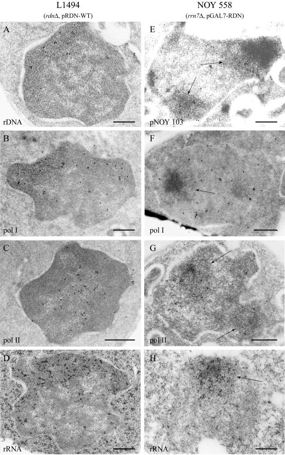
Figure 2.
Electron microscopic localization of the plasmids, RNA pol I, RNA pol II, and 35S rRNA in L1494 and NOY 558. The plasmids bearing rDNA were detected by ISH in the nucleolar regions of both strains L1494 (rdnΔ, pRDN-wt; A) and NOY 558 (rrn7Δ, pGAL7-RDN; E). In NOY 558, the plasmids were precisely localized to the peripheral component. When detected by immunolabeling in L1494, RNA pol I was mainly visible in the nucleolar region (B), whereas pol II was excluded from it (C). In NOY 558, both RNA pol I and RNA pol II were localized to the nucleoplasm and the mininucleoli, the dense component excepted (F and G). In both mutants, ribosomal transcripts were concentrated in the nucleolar regions (D and H). The cytoplasmic labeling corresponds to the ribosomes. In NOY 558, the mininucleoli are designated by arrows. Bar, 300 nm.

