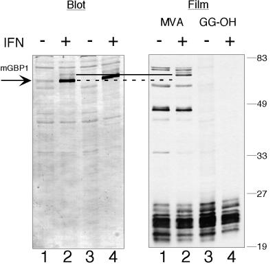
Figure 1.
Incorporation of [3H]isoprenoid into mGBP1 cannot be detected in IFNγ-treated RAW264.7 cells. RAW264.7 cells were labeled for 18 h with either [3H]MVA (lanes 1 and 2) or [3H]GG-OH (lanes 3 and 4) in the absence (lanes 1 and 3) or presence (lanes 2 and 4) of IFNγ and LPS. Total cell lysates were displayed by SDS-PAGE, and mGBP1 was detected by immunoblotting (Blot). “Film” indicates the fluorogram from the immunoblot shown, after 14 d of exposure. With either labeled precursor, no radiolabeled band of the correct size for the induced mGBP1 protein (dotted line) was detected after IFNγ/LPS. However, two new [3H]MVA-labeled proteins (solid line) of slightly slower mobility appeared after IFNγ/LPS treatment. Positions of molecular weight markers are shown on the right.

