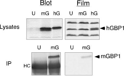
Figure 2.
Poor incorporation of [3H]MVA occurs in cloned mGBP1 expressed in COS-1 cells. (Top) COS-1 cells were labeled for 18.5 h with [3H]MVA in the presence of 25 μM compactin, starting at 30 h after transfection with no DNA (U) or DNAs for mGBP1 (mG) or hGBP1 (hG). Cell lysates were resolved by SDS-PAGE, and radiolabeled proteins were detected by fluorographic exposure for 13 d. (Bottom) After transfection and labeling as described above, mGBP1 was isolated by immunoprecipitation (IP) and separated by SDS-PAGE. Incorporation of radiolabel into mGBP1 was detected after fluorographic exposure for 21 d. “HC” denotes the heavy chain of the anti-GBP antibody.

