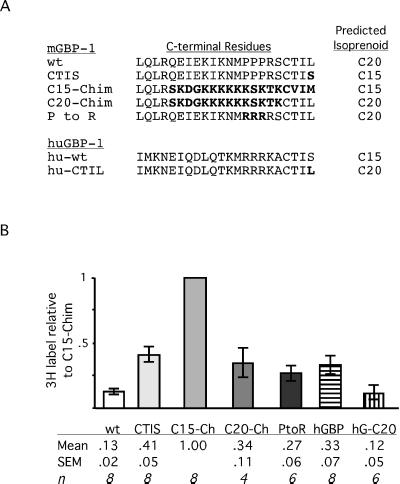
Figure 6.
C-terminal residues of GBPs and [3H]MVA incorporation relative to C15-Chim. (A) C-terminal amino acids of various GBP constructs are aligned. Changes from wild-type mGBP1 or hGBP1 are indicated in boldface type. (B) With the use of a fluorogram and immunoblot from the same membrane, [3H]MVA incorporation and protein amounts of each immunoprecipitated GBP were quantified by scanning, and values of [3H]MVA-derived label were corrected for variations in protein recovery. The amount of 3H for each GBP was then expressed relative to C15-Chim. Values shown are averages of these relative amounts ± SEM. The number of independent experiments is indicated by n. One-way analysis of variance indicated that all GBP proteins incorporated significantly greater amounts of 3H than mGBP1 (p < 0.05), with the exception of the C20-modified hGBP-CTIL (hG-C20), which was not significantly different from mGBP1.

