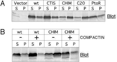
Figure 7.
Membrane association of GBPs that differ in prenylation state. (A) COS-1 cells were transfected with DNAs for the indicated proteins and 48 h later separated into cytosolic (S) and membrane (P) fractions. Samples were separated by SDS-PAGE, and GBPs were detected by immunoblotting. (B) COS-1 cells were transfected with DNAs encoding mGBP1wt or C15-Chim. Treatment with 50 μM compactin was started 5 h later. After 48 h, lysates were separated into cytosolic (S) and membrane (P) fractions and separated by SDS-PAGE, and GBPs were detected by immunoblotting.

