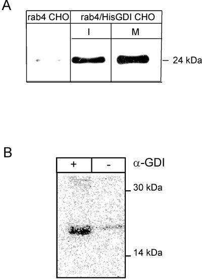
Figure 4.
Released rab4 is not associated with GDI. Equal numbers of interphase (I) and mitotic (M) rab4/HisGDI CHO double transfectants and control rab4 CHO cells were homogenized and fractionated by high speed centrifugation. Cytosol was retrieved and incubated with 50 μl Ni-NTA beads. Beads were washed three times with ice-cold RIPA buffer and boiled in Laemmli sample buffer. Eluted proteins were separated by SDS-PAGE and analyzed by Western blot using the anti Xpress antibody for HisGDI and a rabbit rab4 antibody. Detection was with HRP-labeled secondary antibodies and enhanced chemiluminescence. Quantitation was done using the NIH image software package (A). Mitotic rab4/HisGDI CHO double transfectants were labeled 45 min with 500 μCi/ml 32P ortho phosphate. Cells were lysed in 1% octylglucoside as described in MATERIALS AND METHODS. Equal aliquots cleared lysate were incubated for 1 h with a GDI antibody (+) or preimmune serum (-) adsorbed to Protein A Sepharose CL-4B and washed 3 times with octylglucoside wash buffer. Washed immunoprecipitates were boiled 5 min with 100 μl 0.5% SDS in PBS and pelleted. Supernatants were used to immunoprecipitate rab4 as described inMATERIALS AND METHODS, resolved on 12.5% SDS PAA mini gels, and analyzed by phosphorimaging (B).

