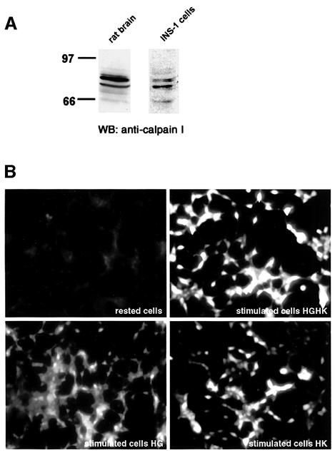
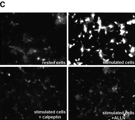
Fig. 3. Expression and activation of µ-calpain in INS-1 cells. (A) Western blotting for µ-calpain I on total protein extracts from rat brain and INS-1 cells. (B) Fluorescence microscopy in live INS-1 cells pre-loaded with the calpain substrate Boc-LM-CMAC and kept in resting conditions or stimulated with 25 mM glucose (HG), 55 mM KCl (HK) or both (HGHK) for 1.5 h. (C) Fluorescence microscopy in live INS-1 cells pre-loaded with the calpain substrate Boc-LM-CMAC and kept in resting conditions or stimulated with HGHK for 1.5 h in the absence or presence of the calpain inhibitors calpeptin and ALLN.

An official website of the United States government
Here's how you know
Official websites use .gov
A
.gov website belongs to an official
government organization in the United States.
Secure .gov websites use HTTPS
A lock (
) or https:// means you've safely
connected to the .gov website. Share sensitive
information only on official, secure websites.

