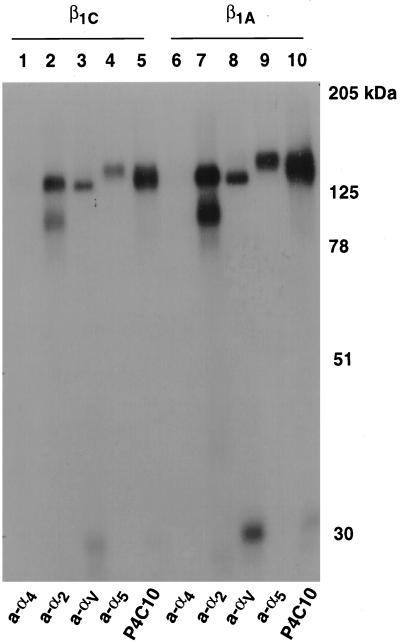
Figure 2.
β1C associates with α5, αV, and α2 subunits. β1C or β1A CHO stable cell lines were cultured for 72 h in the absence of tetracycline and surface-labeled with iodine, and exogenous β1 integrins were immunoprecipitated with P4C10 (lanes 5 and 10). The immunoprecipitated material was then eluted from protein A–Sepharose with 10 mM Tris-HCl, pH 7.5, 0.5% SDS for 10 min at 70°C, reprecipitated with rabbit antiserum to α4 (lanes 1 and 6), α2 (lanes 2 and 7), αV (lanes 3 and 8), or α5 (lanes 4 and 9), and separated by 10% SDS-PAGE. Lanes 1–5, β1C CHO; lanes 6–10, β1A CHO. Proteins were detected by autoradiography. Prestained marker proteins (in kilodaltons) are shown.

