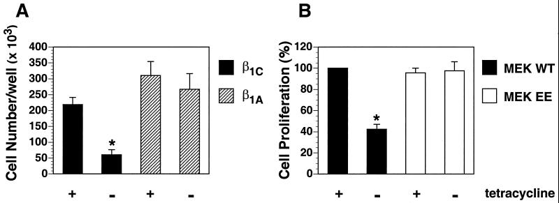
Figure 8.
Constitutively activated MEK rescues β1C inhibition of CHO cell proliferation. (A) β1C or β1A CHO stable cell lines were cultured as described for Figure 7A. Cells were detached, resuspended in serum-free medium, and plated (10,000 cells/well) on tissue culture plates coated with 1 μg/ml fibronectin for 1 h at 37°C. Attached cells were cultured for 96 h at 37°C in growth medium containing 5% FCS either in the absence or in the presence of 1 μg/ml tetracycline. Cells were washed, fixed with 3% paraformaldehyde, and stained overnight with 0.5% toluidine blue. Cell number was evaluated as described in MATERIALS AND METHODS. (B) β1C CHO stable cell lines were transiently transfected with either MEK WT or MEK EE. Cells were cultured for 48 h either in the absence or in the presence of 1 μg/ml tetracycline and starved during the last 24 h of the 48-h culture. Cells were detached, plated, and cultured for 72 h, and cell number was analyzed as described for A. Cell proliferation is expressed as percent relative to the value for MEK WT cultured in the presence of tetracycline. Shown is the average ± SEM from two separate experiments. Group differences were compared with one-way analysis of variance. In A, the differences in proliferation either between β1C CHO stable cell lines in the absence (*) and in the presence of tetracycline or between β1C CHO stable cell lines cultured in the absence of tetracycline (*) and β1A CHO stable cell lines cultured either in the presence or in the absence of tetracycline are statistically significant (p < 0.05). In B, the differences in cell proliferation either between MEK WT in the absence of tetracycline (*) and in the presence of tetracycline or between MEK WT in the absence of tetracycline (*) and MEK EE cultured either in the presence or in the absence of tetracycline are statistically significant (p < 0.05).

