
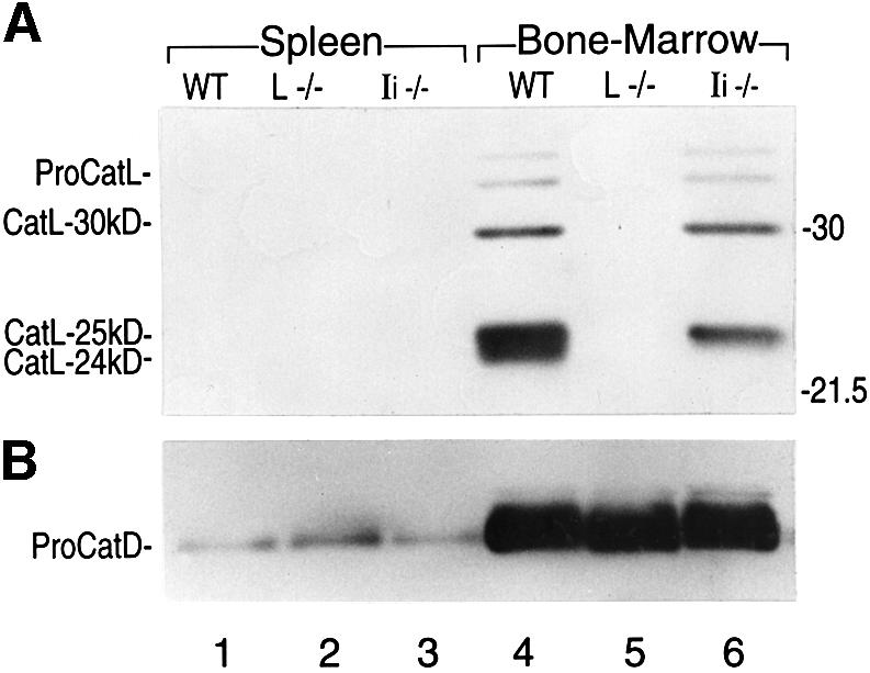
Fig. 1. Expression of CatL in splenocytes and BM APCs from WT and Ii–/– mice. Immunoblot analysis performed on 15 µg of cell lysate obtained from splenocytes or BM cells cultured during 6 days in GM-CSF, and analyzed by SDS–PAGE on a 15% gel under reducing conditions. An antiserum recognizing the proform and the mature forms of CatL (A), or an antiserum raised against CatD (B) was used.