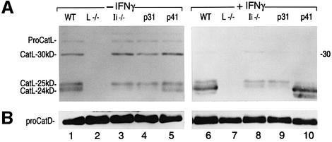
Fig. 2. Expression of CatL in resting and IFNγ-stimulated BM macrophages from WT, Ii–/–, p31 and p41 mice. Immunoblot analysis performed on 15 µg of cell lysate obtained from day 6 BM macrophages cultured for 48 h in the absence (–) or presence (+) of IFNγ, and analyzed by SDS–PAGE on a 15% gel under reducing conditions. An antiserum recognizing the proform and the mature forms of CatL (A), or an antiserum raised against CatD (B) was used. p31 and p41 mice selectively express one Ii isoform.

An official website of the United States government
Here's how you know
Official websites use .gov
A
.gov website belongs to an official
government organization in the United States.
Secure .gov websites use HTTPS
A lock (
) or https:// means you've safely
connected to the .gov website. Share sensitive
information only on official, secure websites.
