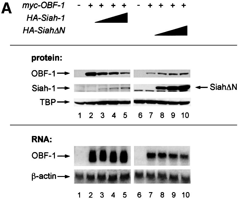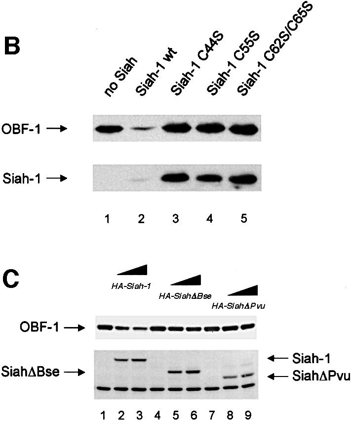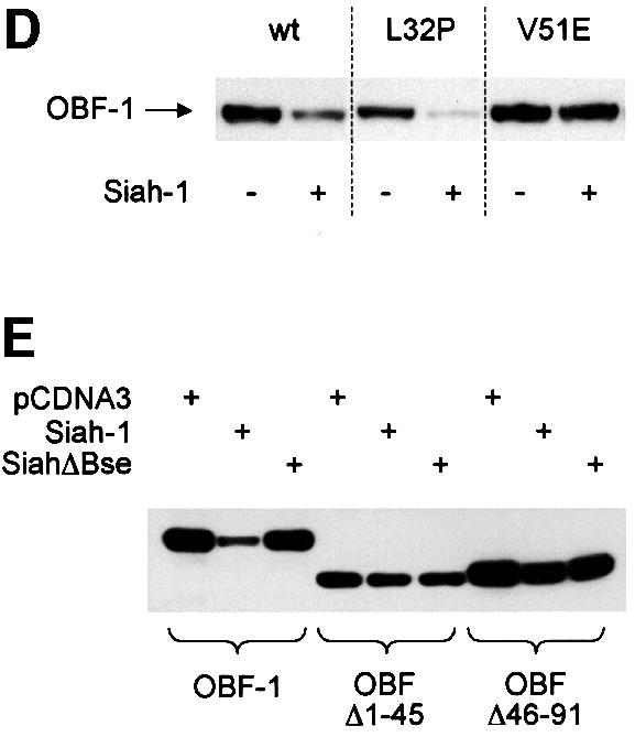


Fig. 3. Interaction between Siah-1 and OBF-1 reduces OBF-1 protein expression. (A) 293T cells were transiently transfected with expression vectors for HA-tagged Siah-1 or Siah-ΔN, and Myc-tagged OBF-1, as indicated. Control transfections in lanes 1 and 6 contained empty expression vectors. Protein extracts and RNA were prepared and equal amounts were analyzed by western (upper panels) or northern blotting (lower panels). OBF-1 and Siah proteins were detected with monoclonal antibodies directed against the respective tags (anti-Myc, 9E10; anti-HA, 12CA5). As a control for endogenous protein, TBP was detected with the specific monoclonal antibody SL-39. For the experiment shown on the right of the figure, less OBF-1 expression vector was used, explaining the weaker signal for OBF-1 protein and RNA. (B) Cells were transfected with Myc-tagged OBF-1 (lanes 1–5) and WT or point mutated Siah (HA-tagged at the N-terminus), as indicated. After protein extract preparation, the respective proteins were detected as described above. Because the Siah-1 point mutant was expressed at a much higher level than WT Siah-1, less expression vector was used for the transfection. (C) Cells were transfected with Myc-tagged OBF-1 (lanes 1–9) and the indicated HA-Siah expression plasmids. OBF-1 and Siah proteins were detected as in (A). The band of equal intensity visible across the lower part of the picture corresponds to an endogenous protein cross-reacting with the anti-HA antibody. (D) Cells were transfected with Myc-tagged expression vectors for WT OBF-1 or the two point mutants indicated, together with a plasmid expressing Siah-1 (lanes 2, 4 and 6). Protein extracts were prepared and OBF-1 protein was detected by western blotting as above. (E) Cells were transfected with Myc-tagged expression vectors for WT OBF-1 or the two deletion mutants indicated, together with a plasmid expressing Siah-1 or SiahΔBse. The level of OBF-1 expression was monitored by western blotting, as above.
