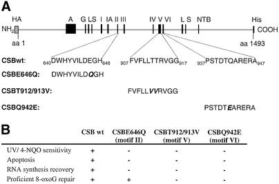
Figure 1.
Diagram showing the CSB protein. (A) The recombinant CSB protein contains a HA epitope; a highly acidic region, A; a glycine rich region, G; two serine phosphorylation signals, S; preceded by NLSs, L; the seven conserved Swi/Snf ATPase motifs, I, IA, II–VI; a NTB fold, NTB; and finally the C-terminal histidine tag, His. Introduced N- and C-terminal tags are shown in gray. Site-directed mutations in ATPase motifs II, V and VI are indicated. (B) Complementation characteristics of the wt and mutant proteins in CS1AN.S3.G2 cells (13,14,18).

