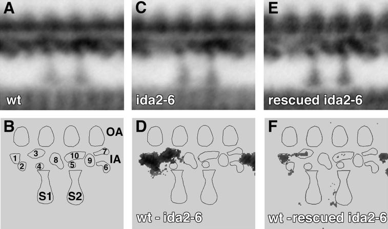
Figure 2.
Inner arm structure in wild-type and mutant strains. Longitudinal images of the 96-nm axoneme repeat from wild type, ida2-6, and the rescued ida2-6 strain, D11, are shown here. The averages for wild type (A), ida2-6 (C), and the rescued ida2-6 strain (E) are based on 8, 10, and 9 individual axonemes and 74, 87, and 85 axoneme repeats, respectively. (B) Model of the 96-nm axoneme repeat with the major lobes of density in the inner arm region labeled 1–10. The outer arms (OA) are on top, the inner arm (IA) region is below, and the proximal and distal radial spokes are labeled S1 and S2, respectively. (D) Difference plot between wild type and ida2-6. (F) Difference plot between wild type and the rescued ida2-6 strain. The difference plots are derived from a pixel-by-pixel analysis of variance between averages, where statistically significant differences between the averages for two strains are indicated by the gray areas. Differences not significant at the 0.005 confidence level have been set to zero.

