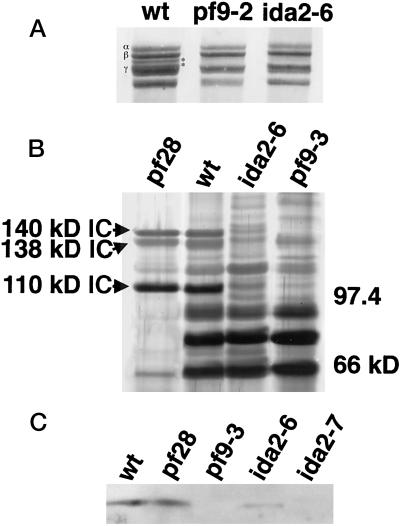
Figure 3.
Dynein polypeptide defects in ida2-6 axonemes. (A) The high-molecular-weight region of a 3–5% polyacrylamide, 3–8 M urea gradient gel that was loaded with whole axonemes from wild type, pf9-2, and ida2-6 is shown here. The 1α and 1β Dhcs of the I1 complex, which migrate between the outer arm β and γ Dhcs, are indicated by the asterisks on the right. Both pf9-2 and ida2-6 lack the 1α and 1β Dhcs. (B) Sucrose density gradient centrifugation of dynein extracts from pf28, wild type, ida2-6, and pf9-3. All sucrose gradient fractions were loaded on 5–15% polyacrylamide gels. The subunits of the I1 complex sediment in the 18–19S region, along with contaminating outer arm components (Piperno et al., 1990; Porter et al., 1992). Only the 18–19S peaks are shown here. IC140, IC138, and IC110 are visible in pf28 (which lacks the outer arm components) and wild type. (C) Western blot of wild-type and mutant axonemes probed with the antibody to the Tctex1 LC.

