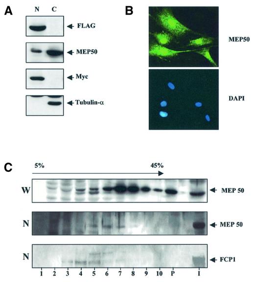
Figure 3.
Subcellular localization of MEP50. (A) HFFCP1 cell fractionation (C, cytoplasmic fraction; N, nuclear fraction) were resolved by SDS–PAGE. Extracts were analyzed by western blotting with anti MEP50 and FLAG and subsequently the same extracts were probed with anti-Myc and anti-tubulin α antibodies as control of the purity and the integrity of nuclear and cytoplasmic protein fractions respectively. (B) MEP50 localize in both nuclear and cytoplasm sub-cellular compartments. An example of immunofluorescence microscopy of human BJ1 cells using MEP50 antibody is reported. DAPI nuclear staining of the same cells is shown. (C) Fractionation of an H1299 cell extract (W, whole cell extract, N, nuclear extract) by ultracentrifugation on a glycerol gradient. Distribution of MEP50 and FCP1 in the gradient fractions and pellet (P) was monitored by western blot using specific antibodies.

