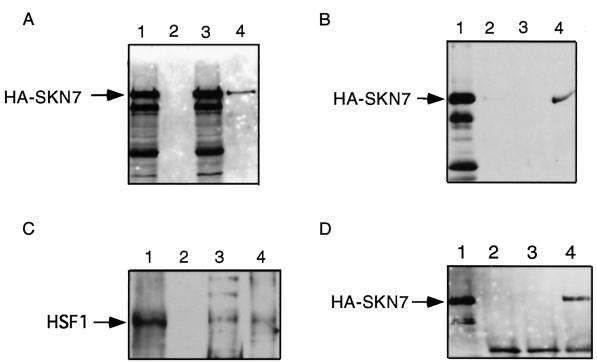
Figure 7.
Hsf1 and Skn7 copurify. (A) Western blot analysis with 12CA5 mAb reveals that pGAL-Skn7-HA copurifies with GST-Hsf1. Lane 1, crude extract containing GST vector alone and pGAL-Skn7-HA; lane 2, GST pull-down from extract containing pGAL-Skn7-HA and GST vector alone; lane 3, crude extract containing pGAL-Skn7-HA and GST-Hsf1; lane 4, GST pull-down from cells containing pGAL-Skn7-HA and GST-Hsf1. (B) Lane 1, 20 μg of input protein (HA-Skn7); lane 2, GST pull-down of extract from cells containing empty GST vector and pGAL-Skn7-HA; lane 3, GST pull-down of extract from cells containing GST-Hsf1 vector and pGAL-Skn7-HA and grown in glucose; lane 4, GST pull-down of extract from galactose-grown cells containing pGAL-Skn7-HA and pGST-Hsf1. (C) Nickel-affinity copurification of Hsf1 with 6His-tagged Skn7. Lane 1, immunoprecipitate with anti-Hsf1 antibody from 1 mg of whole cell extract of galactose-grown cells expressing pGAL-SKN7–6His; lane 2, Ni2+-NTA agarose beads plus 1 mg of cell extract that does not contain the 6His-Skn7 protein; lane 3, Ni2+-NTA agarose beads plus 1 mg of galactose-induced extract from cells expressing pGAL-SKN7–6His; lane 4, 20 μg of input galactose-induced extract. (D) Skn7p can interact with itself. Coimmunoprecipitations from cell extracts containing galactose-induced pGAL-Skn7-HA and 6Myc-Skn7 were performed with the use of 9E10 mAb followed by 12CA5 Western blot analysis. HA-Skn7p expression was under the control of the GAL promoter, and integrated 6Myc-Skn7 was under the control of its own promoter. Immunoprecipitations with 1.5 μg of 9E10 were as follows: lane 1, 20 μg of extract from galactose-grown cells; lane 2, immunoprecipitation of extract from glucose-grown cells; lane 3, immunoprecipitation of extract from galactose-grown cells that did not contain the Myc-tagged SKN7; lane 4; immunoprecipitation of extract from galactose-grown cells containing 6Myc-Skn7 and pGAL-Skn7-HA. Western blot analysis was carried out with 12CA5 mAb.

