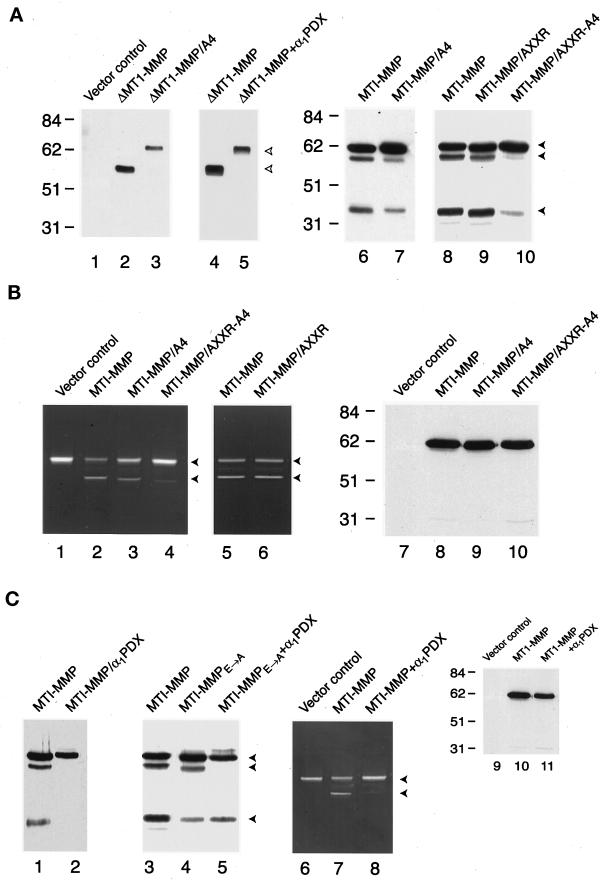
Figure 3.
Processing of membrane-anchored and soluble forms of MT1-MMP. (A) Western blot analysis of MT1-MMP variants harboring mutations in the 108RRKR and/or 86KAMRRPR basic motifs. COS-1 cells were transfected with control (lane 1), ΔMT1-MMP (lanes 2–5), or full-length MT1-MMP (lanes 6–10) expression vectors, and cell-free supernatants (lanes 1–5) or Triton X-114 extracts (lanes 6–10) were analyzed by Western blot analysis with hemopexin domain–specific polyclonal antisera. In lanes 2 and 3, cell-free supernatants of COS-1 cells expressing ΔMT1-MMP or ΔMT1-MMP/A4 were compared, whereas in lanes 4 and 5, the ability of α1PDX to inhibit ΔMT1-MMP processing was assessed. The upper bands in lanes 3 and 5 (marked with open arrowheads) are the pro forms of ΔMT1-MMP. In the case of full-length MT1-MMP (lanes 6–10), COS-1 cells expressing the MT1-MMP/A4 mutant (lane 7) or the MT1-MMP/AXXR-A4 mutant (lane 10) displayed a diminished ability to process the proenzyme relative to cells expressing wild-type MT1-MMP (lanes 6 or 8). The MT1-MMP/AXXR mutant (lane 9) was processed comparably to the MT1-MMP control (lane 8). The closed arrowheads mark the positions of pro, mature, and truncated MT1-MMP. (B) Progelatinase A activation and surface display of MT1-MMP in transiently transfected COS-1 cells. For zymography, COS-1 cells were transiently transfected with control (lane 1), MT1-MMP (lanes 2 and 5), MT1-MMP/A4 (lane 3), MT1-MMP/AXXR-A4 (lane 4), or MT1-MMP/AXXR (lane 6) expression vectors. The COS-1 cells were subsequently incubated with progelatinase A for 16 h, and processing was assessed by zymography (lanes 1–6). The upper and lower arrowheads mark the positions of the pro and fully processed forms of gelatinase A, respectively. In lanes 7–10, control-, MT1-MMP–, or MT1-MMP mutant–transfected COS-1 cells were surface biotinyl-ated, and Triton X-114 extracts were immunoblotted with hemopexin domain–specific polyclonal antisera. (C) Effect of α1PDX on MT1-MMP processing and activity. COS-1 cells were cotransfected with wild-type MT1-MMP and a control expression vector (lanes 1 and 3), MT1-MMPE → A and a control expression vector (lane 4), wild-type MT1-MMP and α1PDX (lane 2), or MT1-MMPE → A and α1PDX (lane 5) expression vectors, and Triton X-114 extracts were analyzed by immunoblotting. Progelatinase A activation was monitored by zymography after incubation with COS-1 cells transfected with the control expression vector (lane 6), with MT1-MMP and a control expression vector (lane 7), or with MT1-MMP and α1PDX expression vectors (lane 8). Cell surface biotinylation followed by Western blot analysis demonstrated that trafficking of proMT1-MMP (lane 10) was not affected by the α1PDX expression vector (lane 11).

