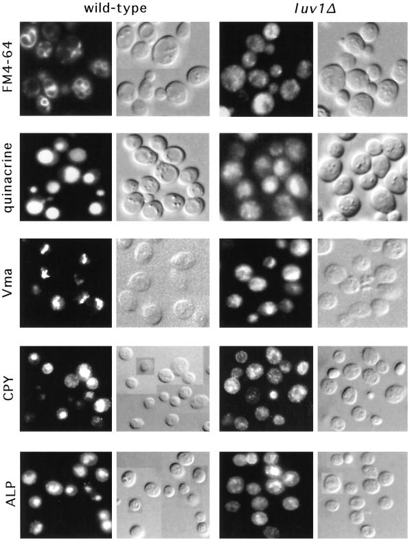
Figure 3.
luv1 mutants accumulate intracellular vesicles that are acidic and contain Vma2p, ALP, and CPY. Wild-type (YPH499) and luv1Δ (YMC1) cells were examined for vacuole morphology and characteristic vacuole proteins. Fluorescence and Nomarski differential interference contrast images are shown. FM4-64 indicates live cells labeled with this membrane dye to show vacuoles. Quinacrine indicates live cells loaded with this dye. Vma, CPY, and ALP indicate fixed cells in which the 60-kDa Vma2p subunit, CPY, and ALP, respectively, were localized by indirect immunofluorescence (see MATERIALS AND METHODS).

