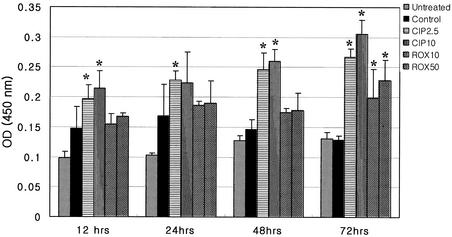
FIG. 2.
DNA fragmentation of activated Jurkat T cells, assessed by ELISA from 12 to 72 h after CPFX or RXM treatment. The results are presented as means ± SEM of absorbance for quadruplicate measurements. Asterisks indicate a P value of <0.05 compared to that of anti-CD3 antibody-treated Jurkat T cells. OD (450 nm), optical density at 450 nm.

