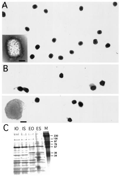
Figure 1.
Negative staining EM and silver-stained gels of OptiPrep and sucrose-purified IMV and EEV preparations. OptiPrep-purified IMV (A) and EEV (B) preparations were absorbed to formvar/carbon-coated grids and were stained with 2% ammonium molybdate. The insets at the bottom left show labeling with antibodies to surface antigens of the IMV (p14 gene A27L) in (A) and EEV (p42; gene B5R) in (B). The bar in the insets is 100 nm. Note that during negative staining using ammonium molybdate the EEV-specific membrane in B tends to collapse on the grid. In (C) sucrose-purified and OptiPrep gradient-purified IMV and EEV preparations were run on 15% SDS-PAGE, and the proteins were detected by silver staining. IO, OptiPrep-purified IMV; IS, sucrose-purified IMV; EO, optiprep-EEV; ES, sucrose-purified EEV; M, marker.

