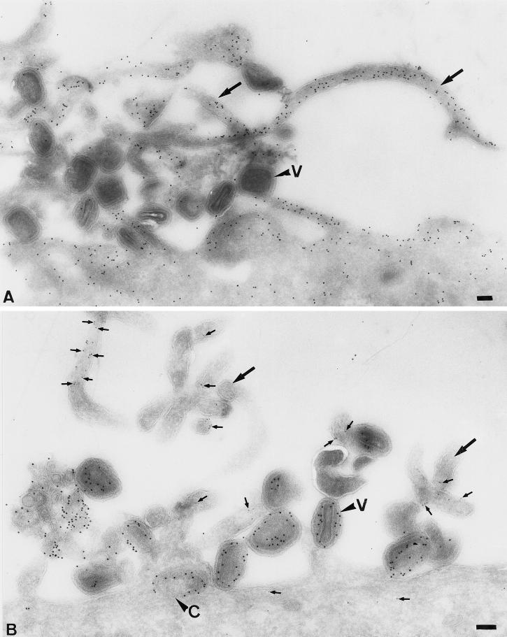
Figure 3.
The exposure of HeLa cells to IMV results in the formation of long microvillar-like structures that contain actin and ezrin. HeLa cells were incubated for 30 min at 37°C with IMV at an MOI of 200. The cells were fixed, prepared for cryosectioning, and labeled with antibodies to actin (A) or were double labeled (B) with antiezrin (5-nm gold, small arrows) and anticore antibody (10-nm gold). Large arrows indicate typical IMV-induced protrusions. V, virions; C, core; bars, 100 nm.

