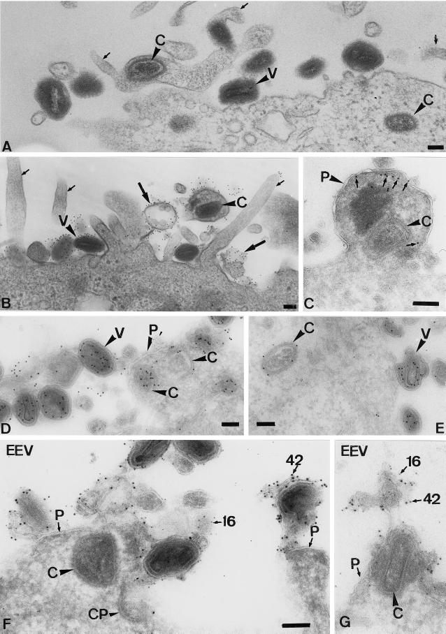
Figure 6.
On entry, the IMV and EEV cores end up “free” in the cytoplasm, while the viral membranes remain at the plasma membrane. (A)–(E) HeLa cells were infected for 30 min at an MOI of 200 with IMV. (A) An Epon-embedded sample with intact virions (V) at the plasma membrane and cores (C) that are free in the cytoplasm. The small arrows indicate viral-induced projections; in one of those projections, a core can be seen that has apparently entered at the level of the microvillus. (B) Epon embedding after preembedding labeling with an antibody to the surface of the IMV. It shows several intact virions that label heavily. Small arrows indicate the microvilli induced by the virus. In the top right, a core has entered a microvillus (which seems to have enlarged), and material adjacent to the surrounding plasma membrane now heavily labels for the antibody to the viral membrane. The image also shows examples of labeled viral membranes (large arrows) devoid of cores that are not connected to the plasma membrane. (C)–(E) Cryosection of IMV-infected HeLa cells. (C) shows a swollen microvillus that contains a core (large arrowhead) that lies free inthe cytoplasm. The small arrows denote ezrin labeling. P, plasma membrane. (D) shows intact virions outside and cores inside the cell both labeled with anticore and clearly separated by the plasma membrane (P). E shows a cytoplasmic core and three virions at the plasma membrane labeled with anticore. The marked virion is clearly changing its morphology. We believe that this change is part of the (elusive) IMV entry mechanism. The section was double labeled with anticore (10-nm gold) and antip16 (5-nm gold). (F) and (G) Cryosections of cells infected with EEV for 10 min. The sections were double labeled for the IMV membrane protein p16 (5-nm gold) and the EEV membrane protein p42 (10-nm gold; both indicated). Whereas the core can be found in the cytoplasm underneath the cell surface, membrane fragments labeled for both p16 and p42 are found attached to the plasma membrane. CP, coated pit. Bars, 100 nm. (F) and (G) are the same magnification.

