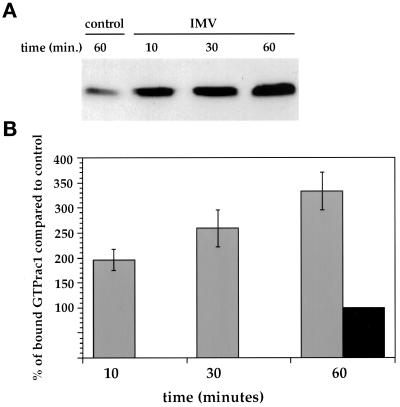
Figure 7.
Exposure of HeLa cells to IMV leads to rac1 activation. (A) shows a representative Western blot of rac1-GTP levels in cell lysates that were either incubated for 60 min in serum-free DMEM (control) or for the indicated times in serum-free DMEM containing purified IMV at an MOI of 40 (IMV). Rac1-GTP was affinity isolated from cell lysates and was detected by ECL. (B) shows the quantitation of four independent experiments. Western blots were probed with antirac1 and antimouse 35SLR, and the blots were quantified by phosphoimager. The average counts of uninfected cells incubated for 60 min in serum-free DMEM (black bar, 60 min) from four experiments was taken as 100%. The gray bars are from HeLa cells exposed to IMV for different periods of time.

