Figure 1.
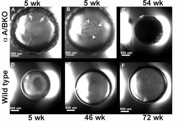
Light micrographs of lenses dissected from fixed eyes of alphaA/BKO (A-C) and wild type (D-F) mice. All micrographs were generated using a 4 × /0.10 objective on a Zeiss LSM 410 using transmitted light mode. Images in (A&B) were enlarged compared to images (C-F) (see scale bars). For all micrographs except (B) the objective focal point was set at the equatorial plane of the lens, while (B) was set at the posterior pole of the lens. (A&B) Representative micrographs of lenses from 5 wk alphaA/BKO mice, showing changes in the equatorial (A) and posterior sub capsular (B) regions. (C) Representative micrograph of 54 wk alphaA/BKO mouse lens showing dense whole lens cataract. (D-F) Representative clear lenses from 5 wk (D), 46 wk (E), and wk 72 (F) wild type mice. Arrows (A) indicate vacuoles in the equatorial region of a representative 5 wk alphaA/BKO lens. The arrowheads (B) indicate an area of minor light scattering deep to the posterior capsule in a representative 5 wk alphaA/BKO lens.

