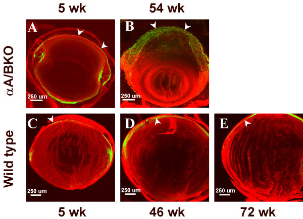
Figure 2.
Representative confocal optical sections taken from mid-axial vibratome sections of alphaA/BKO (A&B) and wild type (C-E) lenses. All optical sections were generated using a 4 × /0.10 objective on a Zeiss LSM 410. The red color represents DiI staining (membrane), while the green color represents SYTOX green staining (DNA). (A&B) were taken from 5 wk and 54 wk alphaA/BKO lenses, respectively. (C-E) were taken from 5 wk, 46 wk, and 72 wk wild type lenses, respectively. The arrowheads indicate representative anterior epithelial nuclei stained with SYTOX green.

