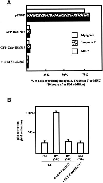
Figure 2.
Rac1 and Cdc42Hs activity and myogenesis. (A) L6 myoblasts were transfected with constructs expressing GFP-tagged Rac1N17 and Cdc42HsN17 and shifted to DM for 50 h. Cells were then fixed and stained for myogenin, troponin T, and MHC expression. L6 myoblasts were also switched to DM containing 10 μM SB 203580. The histogram represents the percentages of myogenin-, troponin T-, or MHC-positive cells and summarizes the data from five independent sets of experiments; 40–50 cells were analyzed in each experiment. (B) L6 myoblasts were cotransfected with HA-p38 and constructs expressing GFP alone as a control, GFP-tagged Rac1N17, or GFP-tagged Cdc42HsN17. p38 activity in the cell lysates was measured as described in MATERIALS AND METHODS from L6 cells in proliferation medium (PM) or L6 cells expressing or not expressing GFP-Rac1N17 or GFP-Cdc42HsN17 in differentiation medium (DM) for 24 h. The results are presented as averages from three independent experiments.

