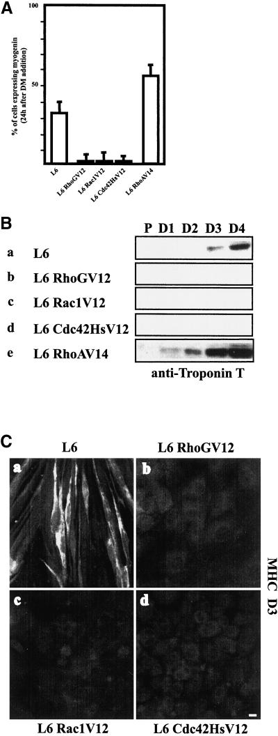
Figure 5.
Effect of RhoGV12, Rac1V12, Cdc42HsV12, and RhoAV14 on muscle-specific protein expression. (A) Proliferating parental L6 and L6 RhoGV12, L6 Rac1V12, L6 Cdc42HsV12, and L6 RhoAV14 myoblasts were grown to ∼80% and shifted to DM for 1 d. Cells were then fixed and stained for myogenin expression and nuclei with Hoechst. The histogram represents the percentage of myogenin-positive cells and summarizes the data from five independent sets of experiments; 40–50 cells were analyzed in each experiment. (B) Protein extracts from parental L6, L6 RhoGV12, L6 Rac1V12, L6 Cdc42HsV12, and L6 RhoAV14 (100 μg/well) collected at the indicated periods (P, proliferative; D1, D2, D3, D4, 1–4 d after addition of differentiation medium) were immunoblotted with an anti-troponin T antibody as described in MATERIALS AND METHODS. Shown are Western blots indicating the expression of troponin T in parental L6 (a), L6 RhoGV12 (b), L6 Rac1V12 (c), L6 Cdc42HsV12 (d), and L6 RhoAV14 (e). (C) Proliferating parental L6 (a) and L6 RhoGV12 (b), L6 Rac1V12 (c), and L6 Cdc42HsV12 (d) myoblasts were grown to ∼80% and shifted to DM for 3 d. Cells were then fixed and stained for MHC expression. For each panel, cells shown are representative of five independent experiments with more than 100 observed cells. Bar, 10 μm.

