Figure 7.
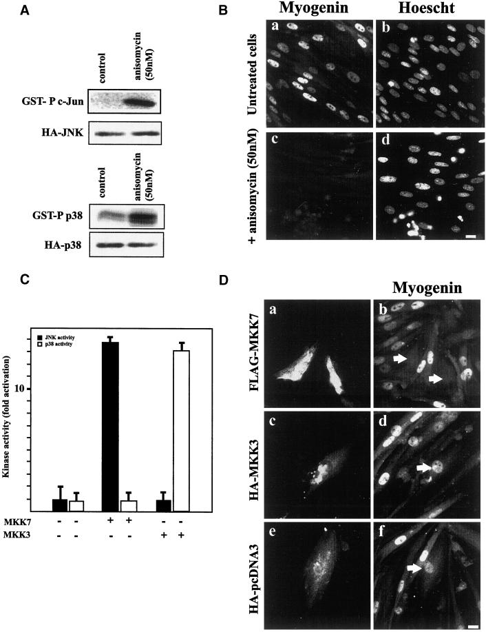
JNK activation impairs myogenin expression. (A) L6 myoblasts were transfected with HA-tagged JNK or HA-tagged p38. Four hours after transfection, cells were treated with 50 ng of anisomycin for 1 d. JNK or p38 activities in the cell lysate were measured by immunocomplex kinase activity with the use of GST–c-jun or GST–ATF2 as a substrate, respectively. HA-JNK and HA-p38 expression were controlled by Western blot analysis with the use of an anti-HA antibody. (B) L6 myoblasts were treated with anisomycin (50 ng) and then induced to differentiate by the addition of DM. One day later, cells were fixed and stained for myogenin expression (a and c) and for DNA with Hoechst dye (b and d). Bar, 10 μm. (C) L6 myoblasts were cotransfected with FLAG-tagged MKK7 or FLAG-tagged MKK3 and either HA-JNK or HA-p38. JNK and p38 activities in the cell lysate were measured by immunocomplex kinase activity with the use of GST–c-jun or GST–ATF2 as a substrate. The results are presented as averages of three independent experiments. (D) L6 myoblasts were transfected with constructs expressing FLAG-MKK7 (a and b), HA-MKK3 (c and d), or empty HA-tagged pcDNA3 (e and f). After transfection, cells were induced to differentiate by the addition of DM, fixed 1.5 d later, and processed for FLAG (a) or HA epitope (c and e) detection and myogenin expression (b, d, and f). For each panel, cells are representative of three independent experiments. Bar, 10 μm.

