
Figure 3.
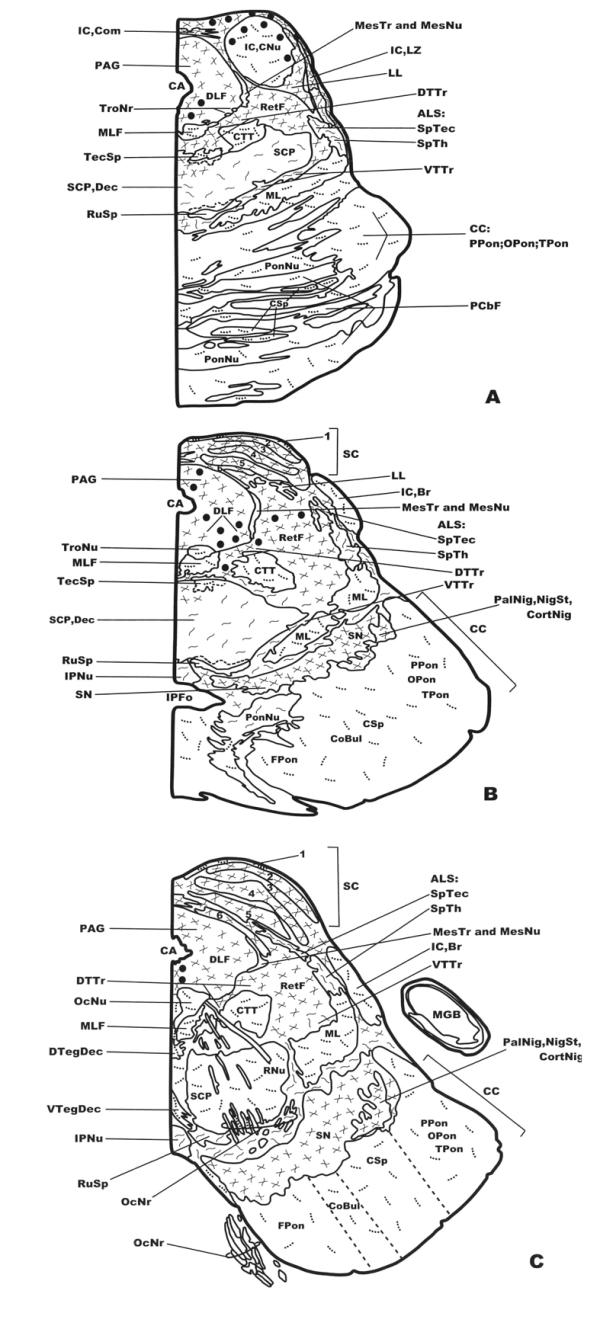
Distribution of NK-ir fibers and cell bodies in frontal planes of the human brainstem from the caudal (Fig. 1A) to the anterior (Fig. 3C) levels. Cell bodies containing neurokinin are represented by closed circles, whereas immunoreactive fibers are represented by dotted lines (single fibers or low density), continuous lines (moderate density) and crossed lines (high density). See list of abbreviations for nomenclature.