Figure 3.
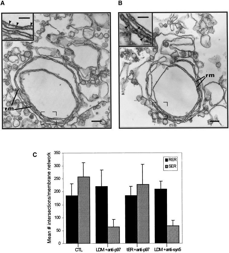
Antibodies to p97 and syntaxin 5 inhibit assembly of smooth tubules in tER. LDM were incubated as for Figure 1b. (a) Antibodies to p97 were present in the incubation medium. (b) Antibodies to syntaxin 5 were present in the incubation medium. (c) Quantitation of p97 and syntaxin 5 antibody effects by morphometry. The amount of rough ER membranes (RER) and smooth endoplasmic reticulum tubules (SER) in reconstituted membrane networks was calculated as indicated in MATERIALS AND METHODS. Microsomes were incubated under control (CTL) conditions as described for Figure 1b or identically in the presence of antibodies to p97 (LDM + anti-p97) or in the presence of antibody to syntaxin 5 (LDM + anti-syn 5). As an additional control, microsomes were preincubated 180 min to promote transitional ER formation and then incubated an additional 60 min in the presence of anti-p97 antibodies (tER + anti-p97). (a and b) Structures labeled rm correspond to parallel rough ER membranes. Insets show high-magnification electron micrographs of parallel rough ER membranes within regions outlined by frames. Scale bars represent 200 nm. (a and b insets) Scale bars represent 100 nm.

