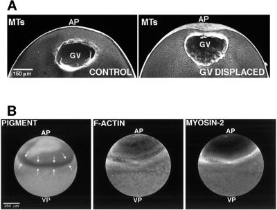
Figure 5.
Redirection of cortical flow by GV displacement. (A) Displacement of the oocyte GV toward the animal pole (AP). Confocal immunofluorescence micrographs of microtubule distribution in a control oocyte and an oocyte subjected to displacement of the GV toward the animal pole before the induction of cortical flow. The GV and its associated microtubules (MTs) are in closer proximity to the animal pole in the oocyte subjected to GV displacement. (B) Cortical pigment, F-actin, and myosin-2 distribution after cortical flow in an oocyte with a displaced GV. Cortical pigment granules, F-actin, and myosin-2 are sparse in the area of the animal pole (AP) toward which the GV was displaced. Instead, they accumulate in a ring positioned between the equator and the animal pole of the oocyte. VP, vegetal pole.

