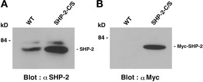
Figure 1.
Expression of SHP-2 in wild-type MDCK cells and MDCK-SHP-2-C/S cells. Cell lysates were prepared from wild-type MDCK cells (WT) and MDCK-SHP-2-C/S cells (SHP-2-C/S) and subjected to immunoblot analysis with the polyclonal anti-SHP-2 Ab (α SHP-2) (A) or the anti-Myc mAb (α Myc) (B). The positions of SHP-2 and Myc-tagged SHP-2 are indicated by bars, and the molecular mass is indicated in kilodaltons (kD).

