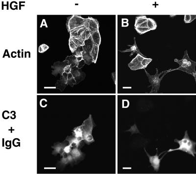
Figure 4.
Reversal by C3 of the inhibition of HGF/SF-induced cell scattering in MDCK-SHP-2-C/S cells. MDCK-SHP-2-C/S cells were microinjected with 40 μg/ml C3 plus 5 mg/ml rat immunoglobulin G (IgG). At 30 min after microinjection, the cells were stimulated with no HGF/SF (A and C) or 10 ng/ml HGF/SF (B and D). At 18 h after HGF/SF stimulation, the cells were fixed, stained with rhodamine-conjugated phalloidin (A and B), and analyzed by confocal microscopy. The microinjected cells are shown by the staining of microinjected rat immunoglobulin G (C and D). Confocal images are shown at the basal level. The results shown are representative of three independent experiments. Bars, 10 μm.

