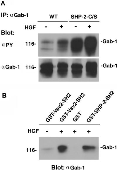
Figure 9.
Tyrosine phosphorylation of Gab-1 and the binding of the SH2 domain of Vav2 to Gab-1. (A) Wild-type cells and MDCK-SHP-2-C/S cells were stimulated with 20 ng/ml HGF/SF for 5 min. Cell lysates were then prepared from HGF/SF-stimulated and unstimulated wild-type MDCK cells and SHP-2-C/S cells and subjected to immunoprecipitation (IP) with the anti-Gab-1 polyclonal Ab (αGab-1). The immunoprecipitates were fractionated by SDS-PAGE and subjected to immunoblot analysis with the anti-phosphotyrosine mAb (αPY). The same filter was then reprobed with the anti-Gab-1 polyclonal Ab. (B) Cell lysates prepared from HGF/SF-stimulated or unstimulated MDCK cells were also incubated with the GST-SH2 domain of Vav2 (GST-Vav2-SH2), the GST-SH2 domains of SHP-2 fusion proteins (GST-SHP-2-SH2), or GST alone (GST) and immobilized on glutathione–Sepharose beads for 2 h. The beads were then subjected to immunoblotting with the anti-Gab-1 polyclonal Ab (αGab-1). The positions of Gab-1 are indicated by bars, and the molecular mass is indicated in kilodaltons on the left.

