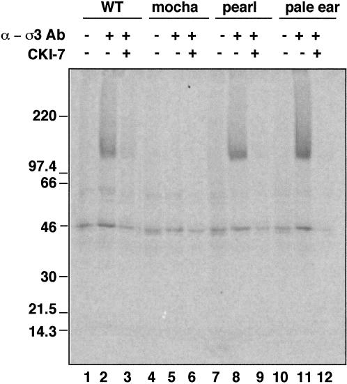
Figure 6.
The neuronal AP-3 complex is associated with casein kinase I. AP-3 complexes from mouse brain cytosol (250 μg/assay) prepared from C57BL6 mice (lanes 1–3), or the Hermansky–Pudlak-like mice mutants mocha (lanes 4–6), pearl (lanes 7–9), and pale ear (lanes 10–12) were immunoprecipitated with preimmune sera (lanes 1, 4, 7, and 10) or antibodies against ς3 subunits. Immunocomplexes were reconstituted at 4°C for 15 min in intracellular buffer in the absence or presence of CKI-7 (250 μM, lanes 3, 6, 9, and 12). Thiophosphorylation was started at 24°C by adding 20 μCi [35S]-ATPγS. Reactions were stopped in ice by adding buffer A plus 20 mM EDTA, were extensively washed, and the complexes were resolved in SDS–PAGE gels. AP-3 complexes immunoprecipitated from pearl mutants that only have the neuronal form of AP-3 were thiophosphorylated. No β3-phosphorylated bands were detected in mocha mice brain cytosol that lacks both neuronal and nonneuronal AP-3 complexes.

