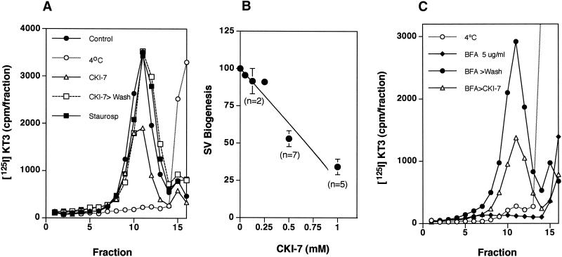
Figure 8.
CKI-7 inhibits in vivo SV budding from PC12 cell endosomes. PC12 N49A cells were labeled with 125I-KT3 at 15°C as described. Unbound radiolabeled antibodies were extensively washed at 4°C, and cells were incubated for 30 min at 15°C in the absence or presence of CKI-7 (500 μM, panel A, or staurosporine (300 nM). In (B) cells were incubated in increasing concentrations of CK1–7. Cells were warmed at 37°C for 20 min to allow SV formation, and the reactions were stopped by chilling down the cells. Recovery experiments (n = 2) were performed after extensively washing the cells with DME media at 4°C before returning them for 20 min to 37°C. Cells were homogenized and fractionated as previously described, and the SV production was assessed in glycerol velocity gradients. CKI-7 inhibited SV budding with an IC50 of 500 μM (B). No inhibition was detected by staurosporine (n = 5). (C) CKI-7 inhibits in vivo endosomal SV budding at a step close to coat recruitment. PC12 N49A cells were labeled with 125I-KT3 at 15°C, unbound label was washed away at 4°C to then incubate the cells either at 4°C (open circles) or for 15 min at 37°C in DME H21 media supplemented with 5 μg/ml brefeldin A to release coat from the membranes. Brefeldin A was kept (closed diamonds) or washed away at 4°C, and the cells were incubated at 15°C for 30 min in the absence (closed circles) or presence of 500 μM CKI-7 (open triangles). Without removing the inhibitor cells, they were warmed at 37°C for 20 min to allow SV formation, and the reactions were stopped by chilling down the cells. Cells were homogenized and fractionated as previously described, and the SV content was assessed in glycerol velocity gradients. CKI-7 hindered SV formation from brefeldin A-treated endosomes as effectively as it inhibited SV formation for endosomes labeled at 15°C. Depicted is a representative experiment of two.

