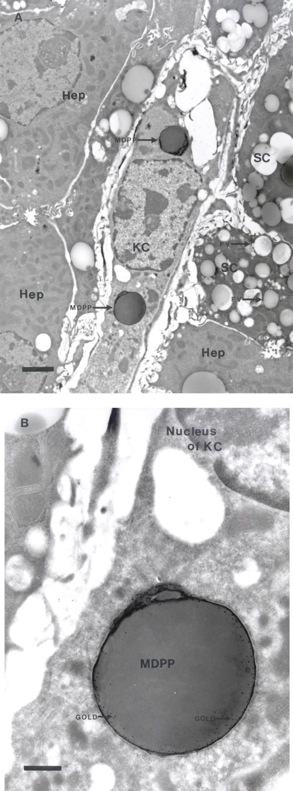
Figure 4.
Uptake of monodisperse polymer particles (MDPP) in Kupffer cells (KC). Following intravenous administration of fluorescently labeled substances, sections were prepared as described in the Methods section for transmission electron microscopy. MDPP are located intracellularly in Kupffer cells, as judged by their characteristic phagocytosis of the particles (A). Hepatocytes (Hep) contain numerous mitochondria. The cells that contain fat vacuoles (FV) may represent stellate cells (SC). To distinguish between vacuoles containing fat and phagocytosed MDPP, sections were immunolabeled with monoclonal anti-mouse TRITC-conjugate. Gold particles are located in the periphery of MDPP where the TRITC-molecules are attached (B). (Scale bars; A: 2 μm, B: 500 nm).

