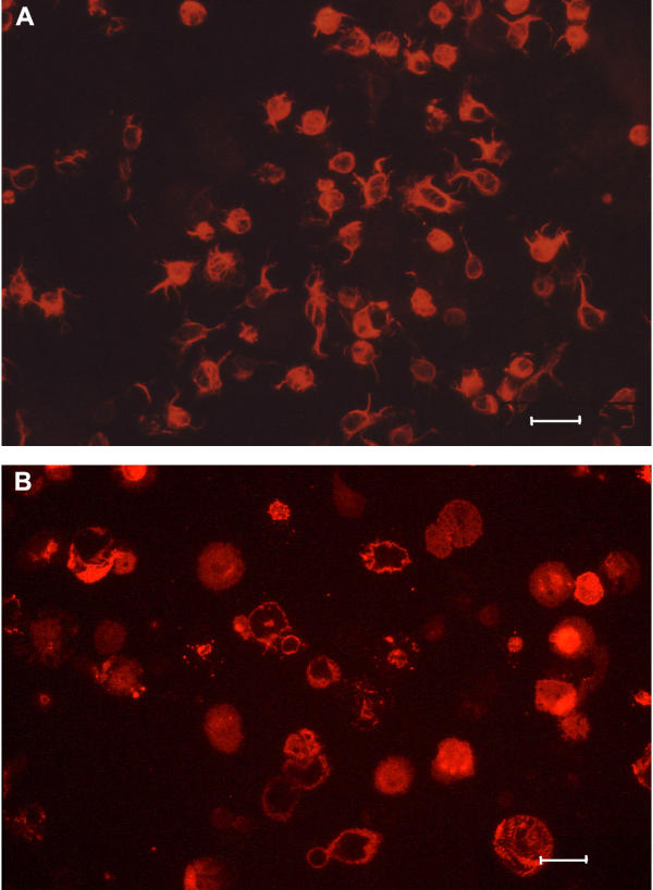
Figure 7.
Fluorescent micrographs of cultured stellate cells and Kupffer cells. Cultures were prepared as described in Methods. The cultures were fixed in 4% paraformaldehyde, after 1 h of incubation. Micrographs of cultured stellate cells stained with monoclonal anti-desmin antibody (A) and cultured Kupffer cells stained with monoclonal anti-pig macrophage antibody (B). (Scale bars; 20 μm).

