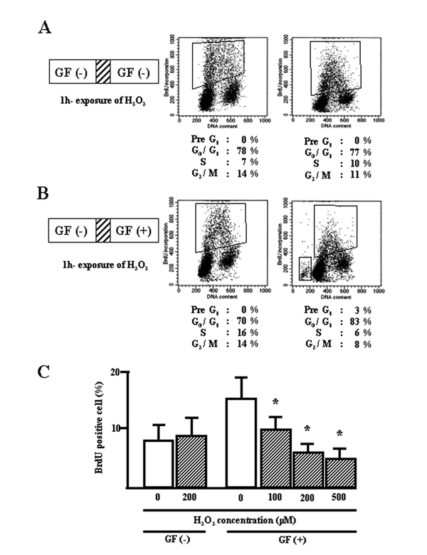
Figure 1.
Distribution of cell cycle phases in BEAS-2B cells exposed to H2O2. A, quiescent cells were exposed to H2O2 for 1 h, followed by culturing in growth factor (GF) free-media for 24 h. B, quiescent cells were exposed to H2O2 for 1 h, followed by culturing in media supplemented with GF for 24 h. All floating and attached cells were collected, incubated with BrdU, and stained with anti-BrdU and PI. C, graphic representation of H2O2-exposed BrdU-positive cells. *, significantly different from control at p < 0.05 (Mann-Whitney U test).

