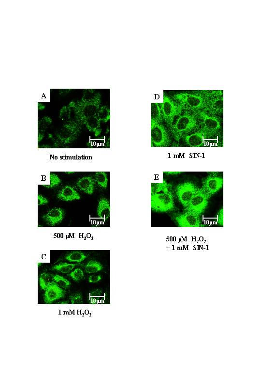
Figure 5.

Indirect immunofluorescence of nitrotyrosine expression in BEAS-2B cells. A, cells without stimulation, B, cells exposed to 500 μM H2O2, C, cells exposed to 1 mM H2O2, D, cells exposed to 1 mM SIN-1, and E, cells exposed to 500 μM H2O2 and 1 mM SIN-1 for 1 h were stained with anti-nitrotyrosine antibody. Each image was captured by laser confocal microscope. Results are representative of 3 individual experiments.
