Figure 2.
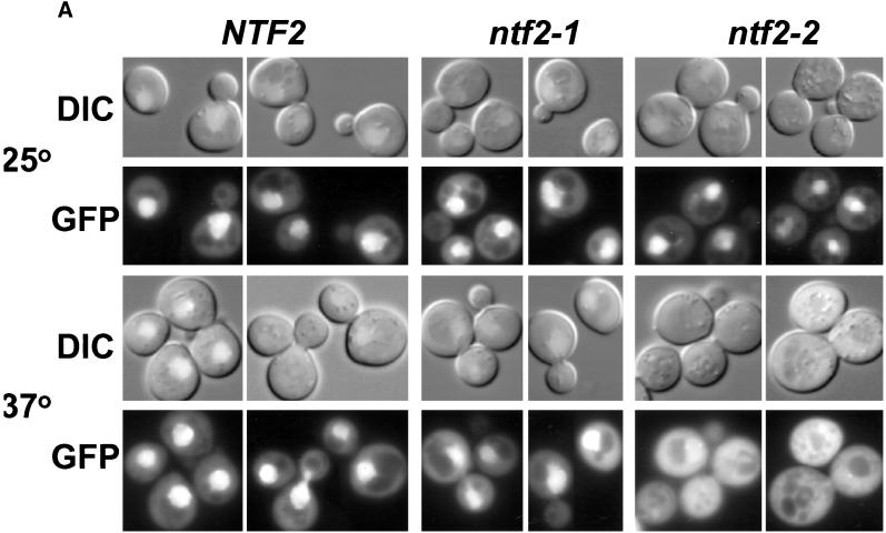
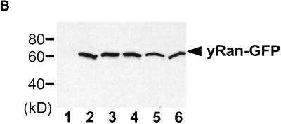
Localization of scRan. (A) NTF2, ntf2–1ts, and ntf2–2ts cells were transformed with a 2μ plasmid encoding scRan-GFP (pAC410). Transformants were grown in liquid media lacking uracil to log phase at 25°C, were split, and were grown at 25°C or 37°C for 3 h. scRan-GFP was viewed directly in living cells. (B) Levels of scRan-GFP are the same in NTF2, ntf2–1ts, and ntf2–2ts transformants grown at 25°C and 37°C. Cells were lysed as described in Materials and Methods, 10 μg of each lysate was resolved on SDS-polyacrylamyide gel, transferred to nitrocellulose, and detected with anti-GFP. Lane 1, vector alone (no GFP) 37°C; lanes 2–4, 25°C (lane 2, NTF2; lane 3, ntf2–1ts; and lane 4, ntf2–2ts); lanes 5 and 6, 37°C (lane 5, ntf2–1ts; lane 6, ntf2–2ts).


